CAR-T cell potency testing with tumour spheroids
Testing the potency of chimeric antigen receptor T (CAR-T) cells using in vitro 3D cell culture models represents a significant advancement in preclinical evaluation methodologies. Traditional 2D monolayer cultures, while informative, fail to recapitulate the complex tumor microenvironment present in vivo. Conversely, 3D cell culture models, such as spheroids, organoids, and scaffold-based systems, provide a more physiologically relevant platform that closely mimics the cellular architecture, heterogeneity, and dynamic interactions within the tumor milieu.
The utilization of patient-derived spheroid models for testing CAR-T cell potency provides a robust, reliable, and scalable platform for the preclinical evaluation of new CAR-T cell therapies. This approach not only accelerates the translational pipeline but also enhances our understanding of CAR-T cell dynamics in a context that closely mirrors the clinical scenario.
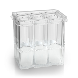
The Sphericalplate 5D® 96w spheroid plate generates 25 spheroids per well, providing 200 spheroids in each removable 8-well strip. This plate design means a high level of convenience and a remarkable improvement in the statistical validity of data, particularly when using patient derived cells. Thanks to the unique round-bottom microwells, cells cluster reliably into consistently sized spheroids. The plate allows for high quality imaging and easy media changes. The Sphericalplate 5D® 96w is pre-coated to prevent cell adhesion and is supplied sterile.

Multiple spheroid replicates per well with SP5D plates
Cat.# 80660M : Expand T-Cells, NK-Cells etc from 5 million / well to 400 million /well with no media change using G-Rex 6M Well Plates.
One of the primary advantages of 3D cultures is their ability to more accurately replicate the intricate characteristics of the tumor microenvironment (TME), which are essential for evaluating CAR-T cell efficacy. The TME encompasses various elements, including hypoxic gradients, nutrient diffusion barriers, extracellular matrix (ECM) components, stromal cells, immune cells, and soluble factors like cytokines and growth factors. These elements collectively influence tumor progression and therapeutic response.
Hypoxic Gradients and Metabolic Constraints: Solid tumors often exhibit regions of low oxygen (hypoxia) due to irregular blood vessel formation and rapid cellular proliferation. Hypoxia can induce a more aggressive phenotype in tumor cells and promote resistance to therapies. 3D culture models can simulate these hypoxic conditions, providing a critical platform for assessing CAR-T cell functionality under stress conditions similar to those in actual tumors.
Nutrient Diffusion Barriers: In vivo tumors experience gradients of nutrient and waste product concentrations due to their three-dimensional structure. These gradients affect cellular metabolism and can create microenvironments where certain cells become more resistant to therapy. 3D models can mimic these diffusion barriers, helping researchers understand how CAR-T cells might perform in nutrient-deprived or acidic conditions, which are common in solid tumors.
Extracellular Matrix (ECM) Composition: The ECM is a network of proteins and glycoproteins that provide structural support to tissues. In tumors, the ECM is often remodeled and can become denser, creating physical barriers to CAR-T cell infiltration. 3D culture systems incorporate ECM components, allowing for the study of CAR-T cell movement and the development of strategies to enhance their penetration through these barriers.
Cellular Heterogeneity: Tumors are composed of a diverse mix of cells, including cancer stem cells, differentiated tumor cells, and various stromal and immune cells. This heterogeneity contributes to treatment resistance and disease recurrence. 3D models allow for the co-culture of different cell types, providing a more comprehensive view of how CAR-T cells interact with and target various cell populations within the tumor.
Stromal and Immune Cell Interactions: The TME includes not only tumor cells but also stromal cells (such as fibroblasts) and immune cells (such as macrophages and regulatory T cells) that can modulate immune responses. These cells can produce immunosuppressive signals that inhibit CAR-T cell activity. By incorporating stromal and immune cells into 3D models, researchers can investigate how these interactions impact CAR-T cell efficacy and explore ways to overcome immunosuppressive barriers.
Cytokine and Growth Factor Gradients: The TME is rich in cytokines and growth factors that influence tumor growth and immune cell behavior. These soluble factors can either stimulate or inhibit CAR-T cell function. 3D models can recreate these gradients, enabling the study of how CAR-T cells respond to various cytokine environments and the identification of key factors that could enhance their activity.